When you need diagnostic or preventive imaging, you want answers you can trust and expert care that puts you at ease. MercyOne Imaging combines advanced technology with experienced imaging professionals to provide high-quality services close to home.

Our team is committed to delivering accurate, timely imaging while making your experience as comfortable and convenient as possible. Patients benefit from:

  • 24/7 imaging coverage to support your care when it’s needed
  • Fellowship-trained radiologists with advanced expertise in interpreting medical images
  • Specially trained and certified technologists who focus on quality, safety and comfort
  • An integrated image storage system that helps your care team securely access and review your images
  • Support for referring physicians to help coordinate care and provide timely results
  • Convenient locations, same-day scheduling options and quick turnaround times.

MercyOne Imaging Exams and Services

Your provider will recommend the imaging exam based on your needs. Services vary by location.

Bone density testing, also called a DEXA scan, measures bone strength and assesses your risk for osteoporosis and fractures. A bone density test can detect bone loss before a fracture occurs and help your provider determine if treatment is needed.  

During the exam, you will lie comfortably on a table while a specialized scanner measures bone density, usually in the hip and spine. The scan is noninvasive and typically takes less than 30 minutes.

Bone density testing may be recommended for:

  • Postmenopausal women
  • Men and women at risk for osteoporosis
  • Patients with a history of fractures
  • Monitoring osteoporosis treatment

Regular bone density testing can help identify bone loss early and support better long-term bone health.

A CT scan, sometimes called a CAT scan, is a quick, painless and noninvasive imaging exam that helps your provider diagnose and monitor many health conditions.

CT technology uses a series of specialized X-ray images and advanced computer processing to create detailed cross-sectional pictures of the inside of your body. These images provide a clearer view of organs, bones, soft tissues and blood vessels than standard X-rays.

CT imaging may be used to help diagnose injuries, evaluate symptoms, guide treatment plans and monitor ongoing conditions. CT services include:

  • Calcium Scoring
  • CT scans
  • Low-dose CT lung screenings
  • Pre-operative scanning

CT perfusion is an imaging test that uses a CT scan and contrast dye to measure blood flow, most often in the brain. It helps doctors identify areas with reduced blood flow, such as during a stroke, and determine which brain tissue may still be saved with treatment.

Fluoroscopy is a specialized imaging exam that uses low-dose X-rays to create real-time images of the inside of your body. Unlike a standard X-ray, fluoroscopy allows your care team to see organs, joints and other structures as they move. This exam helps diagnose a variety of conditions and can also be used to guide certain medical procedures.

Fluoroscopy may also be used during procedures such as joint injections, catheter placement and other image-guided treatments. Some exams require a contrast material to help make specific areas easier to see.

Fluoroscopy is commonly used to evaluate the:

  • Digestive system
  • Urinary tract
  • Joints
  • Reproductive organs

Most fluoroscopy exams require your provider to order the exam and may include special preparation instructions before your appointment.  

Interventional Radiology uses advanced imaging technology to perform minimally invasive procedures that can diagnose and treat a variety of conditions. These procedures are performed by specially trained physicians (interventional radiologists). Because most treatments require only small incisions or needle access, you often experience less discomfort, shorter recovery times and fewer hospital stays compared to traditional surgery.

Interventional Radiology procedures may include:

  • Angiography and Balloon Angioplasty
  • Central Venous Access
  • Gastrostomy Tube Placement
  • Hemodialysis Access Maintenance
  • Needle Biopsy
  • Nephrostomy Tube Placement
  • Thrombolysis
  • Uterine Fibroid Embolization
  • Y-90 Radioembolization 

A mammogram is a special X-ray of the breast that helps detect breast cancer and other breast changes. It is one of the best tools for finding breast cancer early, often before symptoms develop.

During the exam, each breast is gently compressed for a few seconds to create clear images. The exam is quick, and most appointments take less than 30 minutes.

Mammograms are used for routine breast cancer screening and to evaluate concerns such as a lump, breast pain or other changes in the breast.

Schedule your mammogram

Magnetic Resonance Imaging (MRI) uses a powerful magnetic field and radio waves to create highly detailed images of organs, tissues, muscles, joints and other structures inside the body.

Unlike X-rays and CT scans, MRI does not use radiation. MRI is often used to evaluate:

  • Brain and nervous system conditions
  • Joint and sports injuries
  • Spine and back issues
  • Heart and blood vessel conditions
  • Certain cancers and tumors 

Nuclear medicine is a specialized imaging service that helps providers see how organs and tissues are functioning, not just how they look. During the exam, a small amount of radioactive material is given by injection, swallowing or inhalation. Special cameras then capture images that show how your body is working.

Nuclear medicine can help diagnose and monitor many conditions, including:

  • Bone disorders and injuries
  • Certain cancers
  • Gallbladder problems
  • Heart disease
  • Infections
  • Kidney disorders
  • Lung conditions
  • Thyroid conditions

PET/CT combines two advanced imaging technologies into one exam.

  • Positron Emission Tomography (PET) shows how tissues and organs are functioning.
  • Computed Tomography (CT) provides detailed images of the body's structures.

When combined, PET/CT helps providers identify areas of abnormal activity and pinpoint their exact location within the body. PET/CT is commonly used to:

  • Detect and monitor cancer
  • Evaluate treatment response
  • Identify disease recurrence
  • Assist with diagnosis and staging 

Ultrasound, also known as sonography, uses sound waves to create real-time images of structures inside the body. Ultrasound doesn’t use radiation and is a noninvasive imaging option for many patients. Ultrasound can be used to examine:

  • Abdominal aorta and major blood vessels
  • Bladder
  • Breast
  • Gallbladder
  • Infant hips and spine
  • Kidneys
  • Liver
  • Ovaries and uterus
  • Pancreas
  • Prostate
  • Spleen
  • Testicles
  • Thyroid and parathyroid glands 

Vascular screenings are medical tests that check the health of your blood vessels, including arteries and veins. Blood vessels carry blood throughout the body, delivering oxygen and nutrients to organs and tissues. By finding problems early, vascular screenings can help prevent serious conditions like heart attacks, strokes and circulation problems.

Vascular screenings help doctors find signs of issues such as:

  • Blocked arteries that can reduce blood flow
  • Peripheral artery disease (PAD), which affects blood flow to the legs
  • Carotid artery disease, where arteries in the neck become narrowed and may increase stroke risk
  • Abdominal aortic aneurysm (AAA), a dangerous bulging of the body's largest artery 

Whole Body Composition testing provides a detailed look at your body's overall health by measuring body fat, lean muscle mass and bone mass. Unlike a traditional scale, which only shows your total weight, body composition testing helps you understand what your weight is made of. This information can help you track fitness goals, monitor health risks and make informed decisions about nutrition and exercise.

Whole body composition testing can be helpful for:

  • Tracking fitness and wellness goals
  • Monitoring changes in muscle and body fat
  • Supporting weight management programs
  • Evaluating overall health and body composition 

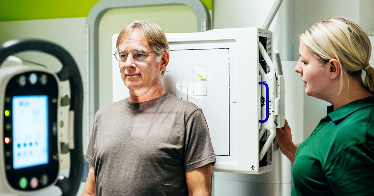
X-ray imaging is one of the most common diagnostic imaging services. It’s used to create images of your bones, lungs and other structures inside your body. X-rays help providers diagnose injuries, illnesses and other medical conditions quickly and accurately.

X-rays are commonly used to evaluate:

  • Broken bones
  • Joint injuries and arthritis
  • Chest and lung conditions
  • Spine and neck concerns
  • Certain abdominal conditions 

At MercyOne, you receive high-quality diagnostic and interventional imaging services delivered with compassion, expertise and efficiency. Our radiologists, technologists and care teams work together to provide an exceptional experience so you receive the answers you need.

  • Convenient access: Many imaging services are available at MercyOne medical centers and care locations across the state, making it easier to receive care close to home.
  • Advanced imaging technology: Our imaging services use the latest technology supported by a system-wide integrated digital image storage system and voice recognition capabilities.
  • Specialized physician expertise: Many MercyOne radiologists are fellowship-trained and have advanced expertise in specific areas of diagnostic or interventional radiology.
  • Experienced imaging professionals: Our highly trained technologists are certified in their imaging specialties and committed to patient comfort, quality, and safety.
  • Recognized quality standards: Many MercyOne imaging locations are accredited by the American College of Radiology, reflecting a commitment to established standards for imaging quality and patient safety.

Your imaging team will tell you how to prepare for your imaging appointment. Instructions vary by scan type.

  • Bring identification, insurance information and any requested order or prior imaging.
  • Ask whether you should eat or drink before your visit.
  • Tell the team about implanted devices, metal, pregnancy, breastfeeding, allergies, kidney concerns or previous contrast reactions.
  • Ask about assistance, an interpreter, accessibility needs or concerns about enclosed spaces.
  • Do not stop medications unless your care team instructs you to do so. 

After your exam, a radiologist reviews the images and sends a report to the provider who ordered the test. That provider will explain how the findings relate to your symptoms, history and next steps.

Ask when to expect results, whether they will appear in your patient portal and whom to contact if you have questions. A report may contain medical terms that need explanation from your care team. 

Frequently Asked Questions

MercyOne offers a full range of imaging services to help diagnose injuries and disease and support preventive care. Available services may vary by location and can include:

The length of your imaging exam depends on the type of test. Some exams, such as X-rays, may take only a few minutes, while CT scans, MRIs and other specialized scans can take longer. Your care team will let you know what to expect before your appointment and answer any questions about preparation or timing.

Some imaging exams use radiation, while others do not. X-rays, CT scans and bone density tests use low doses of radiation to create images. MRI uses magnetic fields and radio waves rather than ionizing radiation.

If you are pregnant, think you may be pregnant or are breastfeeding, please let your provider and imaging team know before your exam. The safest imaging option depends on your individual situation and the type of exam being ordered. Your care team will work with you to determine the most appropriate imaging approach for you and your baby.

Yes. Medical imaging plays an important role in diagnosing and treating illnesses and injury. MercyOne uses advanced imaging technology and follows established safety standards to help ensure every exam is performed safely.

After your exam, the images are reviewed by a radiologist, a physician specially trained in medical imaging. The radiologist interprets the results and shares a report with your provider, who will discuss the findings and any next steps with you.

In most cases, a health care provider will refer you to imaging for a scan to help diagnose a medical condition. Some preventive screenings, such as mammograms, can be self-scheduled. Some MercyOne facilities offer self-pay imaging services.

Pay My Bill

MercyOne offers a range of imaging services at many locations in Iowa.

  • Medical centers
  • Urgent care locations
  • Imaging centers
  • Primary care locations 

Many diagnostic imaging exams require an order from a health care provider. Requirements depend on the exam, location and insurance plan. Some screening mammograms may be scheduled directly. Contact the imaging location to confirm what is needed before your visit.

Follow the instructions provided by your imaging team. Preparation depends on the exam and may involve eating or drinking instructions, medication guidance or removing metal items. Tell the team about pregnancy, implanted devices, allergies, kidney concerns or previous reactions to contrast. Do not stop medications unless your care team tells you to.

Some exams use contrast material to help show specific tissues or blood vessels. Whether contrast is needed depends on the exam and the clinical question. Tell the imaging team about prior contrast reactions, allergies, kidney concerns, pregnancy or breastfeeding so it can provide instructions specific to you.

Tell your ordering provider and imaging team if you are pregnant, may be pregnant or are breastfeeding before the exam. The appropriate test and any contrast instructions depend on your situation. Your care team will discuss options and determine whether the exam should proceed or be adjusted.

A radiologist interprets the images and sends a report to the clinician who ordered the exam. Ask your care team when to expect results, how they will be shared and whom to contact with questions. Portal release timing and follow-up arrangements may vary.